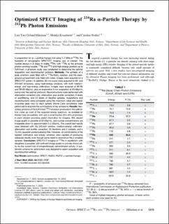
| dc.description.abstract | In preparation for an a-particle therapy trial using 1–7 MBq of 224Ra, the feasibility of tomographic SPECT/CT imaging was of interest. The nuclide decays in 6 steps to stable 208Pb, with 212Pb as the principle photon-emitting nuclide. 212Bi and 208Tl emit high-energy photons up to 2,615 keV. A phantom study was conducted to determine the optimal acquisition and reconstruction protocol. Methods: The spheres of a body phantom were filled with a 224Ra-RaCl2 solution, and the back- ground compartment was filled with water. Images were acquired on a SPECT/CT system. In addition, 30-min scans were acquired for 80- and 240-keV emissions, using triple-energy windows, with both medium- energy and high-energy collimators. Images were acquired at 90–95 and 29–30 kBq/mL, plus an explorative 3-min acquisition at 20 kBq/mL (using only the optimal protocol). Reconstructions were performed with attenuation correction only, attenuation plus scatter correction, 3 levels of postfiltering, and 24 levels of iterative updates. Acquisitions and reconstructions were compared using the maximum value and signal- to-scatter peak ratio for each sphere. Monte Carlo simulations were performed to examine the contributions of key emissions. Results: Sec- ondary photons of the 2,615-keV 208Tl emission produced in the collima- tors make up most of the acquired energy spectrum, as revealed by Monte Carlo simulations, with only a small fraction (3%–6%) of photons in each window providing useful information for imaging. Still, decent image quality is possible at 30 kBq/mL, and nuclide concentrations are imageable down to approximately 2–5 kBq/mL. The overall best results were obtained with the 240-keV window, medium-energy collimator, attenuation and scatter correction, 30 iterations and 2 subsets, and a 12-mm gaussian postprocessing filter. However, all combinations of the applied collimators and energy windows were capable of producing adequate results, even though some failed to reconstruct the 2 smallest spheres. Conclusion: SPECT/CT imaging of 224Ra in equilibrium with daughters is possible, with sufficient image quality to provide clinical util- ity for the current trial of intraperitoneally administrated activity. A sys- tematic scheme for optimization was designed to select acquisition and reconstruction settings. | en_US |

