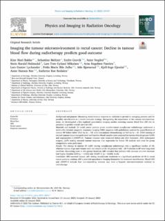
Imaging the tumour microenvironment in rectal cancer: Decline in tumour blood flow during radiotherapy predicts good outcome
| dc.contributor.author | Bakke, Kine Mari | |
| dc.contributor.author | Meltzer, Sebastian | |
| dc.contributor.author | Grøvik, Endre | |
| dc.contributor.author | Negård, Anne | |
| dc.contributor.author | Holmedal, Stein Harald | |
| dc.contributor.author | Mikalsen, Lars Tore Gyland | |
| dc.contributor.author | Færden, Arne Engebreth | |
| dc.contributor.author | Lyckander, Lars Gustav | |
| dc.contributor.author | Julbø, Frida Marie Ihle | |
| dc.contributor.author | Bjørnerud, Atle | |
| dc.contributor.author | Gjesdal, Kjell Inge | |
| dc.contributor.author | Ree, Anne Hansen | |
| dc.contributor.author | Redalen, Kathrine Røe | |
| dc.date.accessioned | 2023-09-08T12:28:40Z | |
| dc.date.available | 2023-09-08T12:28:40Z | |
| dc.date.created | 2023-02-27T13:24:44Z | |
| dc.date.issued | 2023 | |
| dc.identifier.citation | Physics and imaging in radiation oncology (PIRO). 2023, 25:100417 1-7. | en_US |
| dc.identifier.issn | 2405-6316 | |
| dc.identifier.uri | https://hdl.handle.net/11250/3088328 | |
| dc.description.abstract | Background and purpose Measuring rectal tumour response to radiation is pivotal to restaging patients and for possibly stratification to a watch-and-wait strategy. Recognizing the importance of the tumour microenvironment, we investigated a less explored quantitative imaging marker assessing tumour blood flow (BF) for its potential to predict overall survival (OS). Materials and methods 24 rectal cancer patients given curative-intent neoadjuvant radiotherapy underwent a multi-echo dynamic magnetic resonance imaging (MRI) sequence with gadolinium contrast for quantification of tumour BF before either 25x2 Gy (n = 18) with concomitant chemotherapy or 5x5 Gy (n = 6). CD34 staining of excised tumour tissue was performed and baseline blood samples were analysed for lactate dehydrogenase (LDH) and angiopoietin-2 (ANGPT-2). Tumour volumes were measured before and after treatment. After subsequent surgery, ypTN scoring assessed tumour response. Cox regression for 5-year OS analysis and t-test for group comparisons were performed. Results The change in tumour BF (ΔBF) during neoadjuvant radiotherapy was a significant marker of OS, whereas tumour stage and volume were not related to OS. All patients with >20 % decline in BF were long-term survivors. Separating cases in two groups based on ΔBF revealed that patients with increase or a low decrease had higher baseline LDH (p = 0.032) and ANGPT-2 (p = 0.028) levels. Conclusion MRI-assessed tumour ΔBF during neoadjuvant treatment is a significant predictor of OS in rectal cancer patients, making ΔBF a potential quantitative imaging biomarker for treatment stratification. Blood LDH and ANGPT-2 indicate that non-responding tumours may have a hypoxic microenvironment resistant to radiotherapy. | en_US |
| dc.language.iso | eng | en_US |
| dc.relation.uri | https://phiro.science/article/S2405-6316(23)00008-8/fulltext | |
| dc.rights | Navngivelse 4.0 Internasjonal | * |
| dc.rights.uri | http://creativecommons.org/licenses/by/4.0/deed.no | * |
| dc.title | Imaging the tumour microenvironment in rectal cancer: Decline in tumour blood flow during radiotherapy predicts good outcome | en_US |
| dc.type | Peer reviewed | en_US |
| dc.type | Journal article | en_US |
| dc.description.version | publishedVersion | en_US |
| cristin.ispublished | true | |
| cristin.fulltext | original | |
| cristin.qualitycode | 1 | |
| dc.identifier.doi | 10.1016/j.phro.2023.100417 | |
| dc.identifier.cristin | 2129644 | |
| dc.source.journal | Physics and imaging in radiation oncology (PIRO) | en_US |
| dc.source.volume | 25:100417 | en_US |
| dc.source.pagenumber | 1-7 | en_US |
| dc.relation.project | Kreftforeningen: 198116 | en_US |
Tilhørende fil(er)
Denne innførselen finnes i følgende samling(er)
-
HV - Institutt for naturvitenskapelige helsefag [392]
HV - Department of Life Sciences and Health -
Publikasjoner fra Cristin [3201]

Med mindre annet er angitt, så er denne innførselen lisensiert som Navngivelse 4.0 Internasjonal