| dc.contributor.author | Hovda, Tone | |
| dc.contributor.author | Hoff, Solveig Roth | |
| dc.contributor.author | Larsen, Marthe | |
| dc.contributor.author | Romundstad, Linda | |
| dc.contributor.author | Sahlberg, Kristine Kleivi | |
| dc.contributor.author | Hofvind, Solveig | |
| dc.date.accessioned | 2022-12-12T08:43:04Z | |
| dc.date.available | 2022-12-12T08:43:04Z | |
| dc.date.created | 2021-11-01T13:46:37Z | |
| dc.date.issued | 2021-04-21 | |
| dc.identifier.citation | Academic Radiology. 2021, 29 S180-S191. | en_US |
| dc.identifier.issn | 1076-6332 | |
| dc.identifier.uri | https://hdl.handle.net/11250/3037129 | |
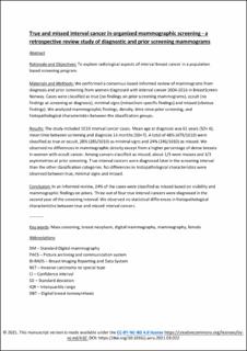
| dc.description.abstract | Rationale and Objectives: To explore radiological aspects of interval breast cancer in a population based screening program.
Materials and Methods: We performed a consensus-based informed review of mammograms from diagnosis and prior screening from women diagnosed with interval cancer 2004-2016 in BreastScreen Norway. Cases were classified as true (no findings on prior screening mammograms), occult (no findings at screening or diagnosis), minimal signs (minor/non-specific findings) and missed (obvious findings). We analyzed mammographic findings, density, time since prior screening, and histopathological characteristics between the classification groups.
Results: The study included 1010 interval cancer cases. Mean age at diagnosis was 61 years (SD= 6), mean time between screening and diagnosis 14 months (SD=7). A total of 48% (479/1010) were classified as true or occult, 28% (285/1010) as minimal signs and 24% (246/1010) as missed. We observed no differences in mammographic density except from a higher percentage of dense breasts in women with occult cancer. Among cancers classified as missed, about 1/3 were masses and 1/3 asymmetries at prior screening. True interval cancers were diagnosed later in the screening interval than the other classification categories. No differences in histopathological characteristics were observed between true, minimal signs and missed.
Conclusion: In an informed review, 24% of the cases were classified as missed based on visibility and mammographic findings on priors. Three out of four true interval cancers were diagnosed in the second year of the screening interval. We observed no statistical differences in histopathological characteristics between true and missed interval cancers. | en_US |
| dc.language.iso | eng | en_US |
| dc.publisher | Elsevier | en_US |
| dc.relation.ispartofseries | Academic Radiology;Volume 29, Supplement 1, January 2022, Pages S180-S191 | |
| dc.rights | Attribution-NonCommercial-NoDerivatives 4.0 Internasjonal | * |
| dc.rights.uri | http://creativecommons.org/licenses/by-nc-nd/4.0/deed.no | * |
| dc.subject | Mass screening | en_US |
| dc.subject | Breast neoplasm | en_US |
| dc.subject | Digital mammography | en_US |
| dc.subject | Mammography | en_US |
| dc.subject | Female | en_US |
| dc.title | True and Missed Interval Cancer in Organized Mammographic Screening: A Retrospective Review Study of Diagnostic and Prior Screening Mammograms | en_US |
| dc.type | Peer reviewed | en_US |
| dc.type | Journal article | en_US |
| dc.description.version | acceptedVersion | en_US |
| cristin.ispublished | true | |
| cristin.fulltext | original | |
| cristin.fulltext | postprint | |
| cristin.qualitycode | 1 | |
| dc.identifier.doi | https://doi.org/10.1016/j.acra.2021.03.022 | |
| dc.identifier.cristin | 1950250 | |
| dc.source.journal | Academic Radiology | en_US |
| dc.source.volume | 29 | en_US |
| dc.source.issue | Supplement 1 | en_US |
| dc.source.pagenumber | S180-S191 | en_US |

